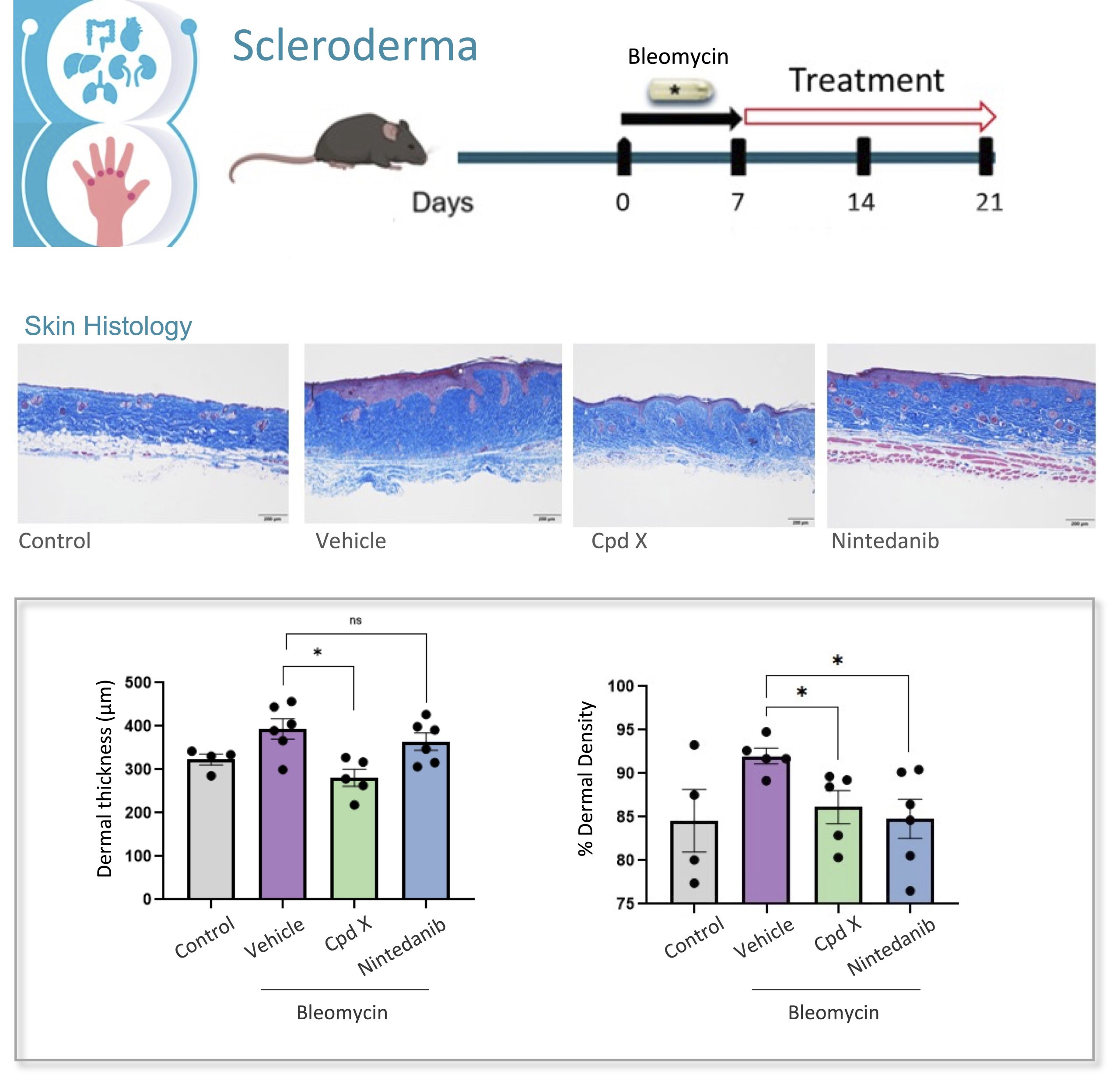
Scleroderma or systemic sclerosis is a rare connective tissue disorder with complex pathogenesis. Scleroderma can be divided in localized scleroderma primarily affecting the skin and subcutaneous tissue, whereas systemic sclerosis is associated with systemic manifestations and involvement of multiple organ systems, including kidney, lung, heart, gastrointestinal tract and more.
To support the development of antifibrotic therapies that will be effective in the treatment of scleroderma we have developed and standardized the bleomycin induced scleroderma animal model, that reproduces the pathology manifestations and is widely use in preclinical research.
The scleroderma model induced by mini osmotic pump released bleomycin integrates comprehensive histopathological and molecular readouts of fibrosis and inflammation, providing a well-established and highly translational in vivo preclinical platform for evaluating novel therapeutics targeting fibrosis.
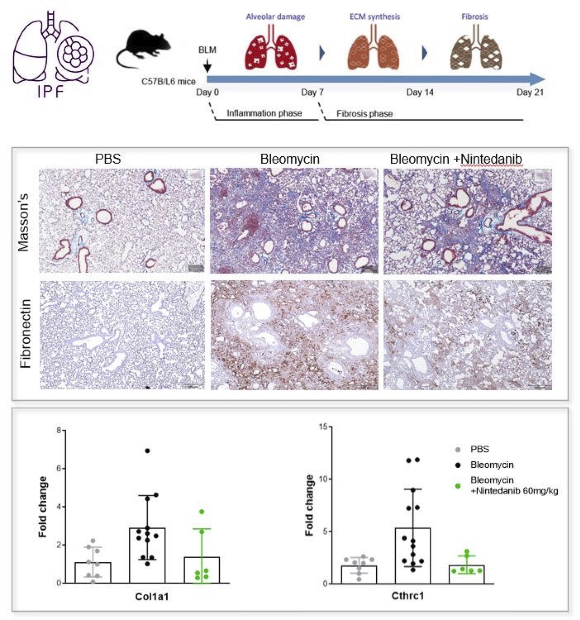
🫁 Idiopathic Pulmonary Fibrosis (IPF) remains one of the most devastating chronic lung diseases, driven by complex dysregulated pathways involving multiple cell types, including macrophages, fibroblasts, and epithelial cells. Characterized by progressive lung inflammation and scarring, IPF continues to present significant challenges for patients due to limited therapeutic options.
💊Advancing effective anti-fibrotic therapies requires reliable and translational preclinical models that can accurately capture disease biology and support robust efficacy evaluation before clinical development.
The bleomycin-induced IPF mouse model developed in Biomedcode integrates comprehensive histopathological and molecular readouts of fibrosis and inflammation, providing a well-established and highly translational in vivo preclinical platform for evaluating novel therapeutics targeting fibrosis.
INFRAFRONTIER has achieved two major milestones in the first half of 2024, with two collaborative projects being funded by the European Commission INFRAPLUS and PRIMTECH3R
Idiopathic pulmonary fibrosis (IPF) is a chronic and progressive interstitial lung disease characterized by irreversible fibrosis. It involves the transition of fibroblasts to myofibroblasts, the tissue stiffening and alveolar epithelium injury eventualy causing irreversible and progressive lung damage.
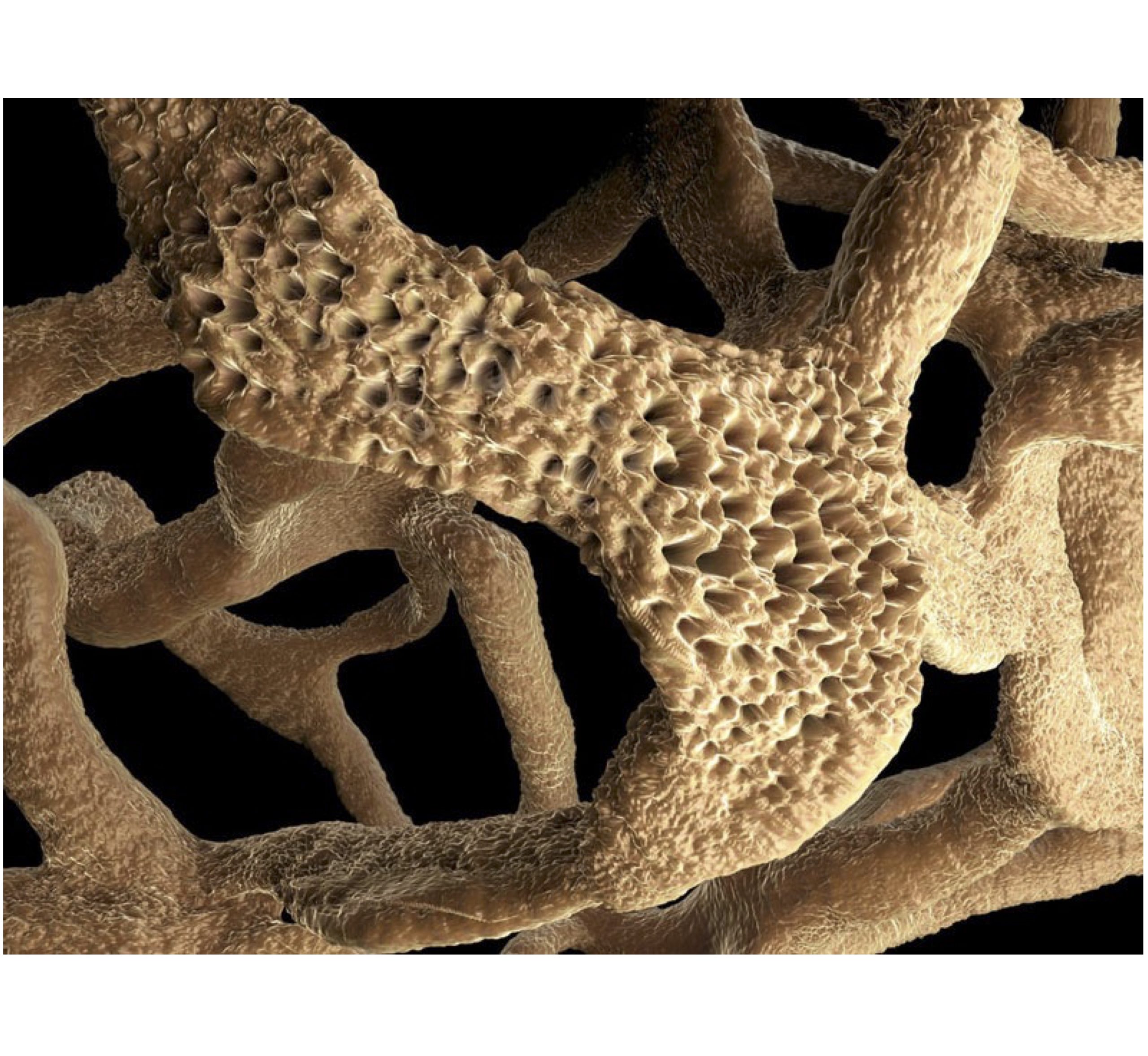
Various methods have been employed to simulate pulmonary fibrosis for the purpose of evaluating potential anti-fibrotic therapeutic treatments. Apart from the use of IPF animal models where the pathology is induced through the endotracheal administration of bleomycin and the development of 2D cell cultures for the study of fibrosis mechanisms, three-dimensional (3D) cultures have also emerged as valuable tools for modeling IPF.
One such 3D culture approach is the generation of precision-cut lung slices (PCLS), where thin slices with precise thickness are obtained from fresh tissue with the use of a vibrating microtome. These slices recapitulate the in vivo lung environment by offering a preserved tissue structure with intact cell-cell and cell-extracellular matrix interactions, allowing for the study of molecular mechanisms between different lung cell populations and facilitating lung degeneration and alveologenesis. The advantage of this approach is that it allows for the generation of multiple slices from the lungs of a single mouse , thereby greatly reducing the number of animals required for individual experiments.
As it has been shown that PCLS obtained from IPF patients or bleomycin-induced mouse lungs, exhibit reduced expression of fibrotic markers following TGF-β inhibition, Biomedcode is currently developing a preclinical ex vivo tool using PCLS derived from bleomycin-induced mouse lungs to screen ex vivo the therapeutic efficacy of anti-fibrotic drugs. Using Nintedanib, an intracellular tyrosine kinase inhibitor that targets fibroblast recruitment, proliferation and differentiation, as a positive control and through the assessment of fibrosis biomarkers such as fibronectin, alpha Sma and collagen 1A1, we have generated experimental evidence supporting that PCLS from IPF-affected mouse lungs can nicely serve as a powerful preclinical tool to assess the anti-fibrotic effect of test articles targeting inflammation and fibrosis.
TNF is a pleiotropic cytokine serving important functions in pathophysiology, as it plays regulatory role in the growth, differentiation and death of immune and non-immune cells. Its functions are mediated through binding to two transmembrane receptors, TNFR1 and TNFR2, that deliver signals to activate a variety of responses ranging from proliferation to apoptosis and necrosis.
Dysregulation of TNF has been implicated in immune mediated inflammatory diseases including rheumatoid arthritis, inflammatory bowel disease, psoriasis, scleroderma, systemic lupus erythematosus, atherosclerosis as well as autoimmune pathologies, cancer etc.
Since the approval of the first TNF inhibitors, back in 1998, TNF blockade has been used as a successful approach in treating a broad range of pathologies, having nonetheless the expected compromises stemming from the severe side effects that result from the blockade of the whole range of the physiological functions of TNF, with the increased susceptibility to infections being one of the majordrawbacks of this therapeutic approach.
The pioneering work of two Greek scientists, Kassiotis and Kollias, published in 2001 in JEM (193:427) and JCI (107:1507), shed light for the first time on the importance ofuncoupling the TNFR1- and TNFR2-TNF signaling pathways. This work proposed that specific blockade of TNFR1 signaling could offer advantageous novel therapeutic approaches as they would suppress the TNF pro-inflammatory activity, leaving unaffected its TNFR2-dependent immunosuppressive activity. In the following years, indeed TNFR1 emerged as an attractive target towards ameliorating the detrimental effects of the TNF signalling pathway and various TNFR1 inhibitors are currently under development or in clinical trials.
A unique tool for testing anti-hTNFR1 therapeutics is the human TNFR1 knock in (hTNFR1KI) mouse model, that expresses human TNFR1 in the absence of its mouse counterpart. The hTNFR1KI mouse combined, as shown in the table below, with induced or spontaneous disease models offers a fully equipped armory for the evaluation of human therapeutics targeting TNFR1.
Preclinical Platforms for the evaluation therapeutics targeting human TNFR1
One of the models described above that has a particular interest due to its complexity and similarity to the human condition is the TNFΔAREhTNFR1KI mouse model that due to the dysregulated expression of TNF develops spontaneously intestinal inflammation, arthritis and cardiovascular disease, perfectly recapitulating many of the features that constitute the complexity of the human disease. TNFΔARE was the first mouse model that unequivocally established the causal role of TNF in the inflammatory response of Crohn’s-like ileitis, while further studies revealed numerous promising therapeutic targets some of which were later developed into approved drugs, such as Ustekinumab, vedolizumab, and natalizumab.
The TNFΔAREhTNFR1KI mouse model, faithfully reproduces all TNFΔARE phenotypes and is an ideal tool for translational research, supporting the drug discovery and development of biologics and small molecules targeting TNF, hTNFR1 as well as other potential therapeutic targets of Crohn’s-like ileitis.
Extending on our previous work on the treatment of arthritis with combination therapies of dasatinib with subtherapeutic doses of anti-TNF biologics, we explore here in this collaborative research project the interplay of inflammation and senescence in the context of combination treatments.
Published in Mechanisms of Ageing and Development2023, 214, 111856.
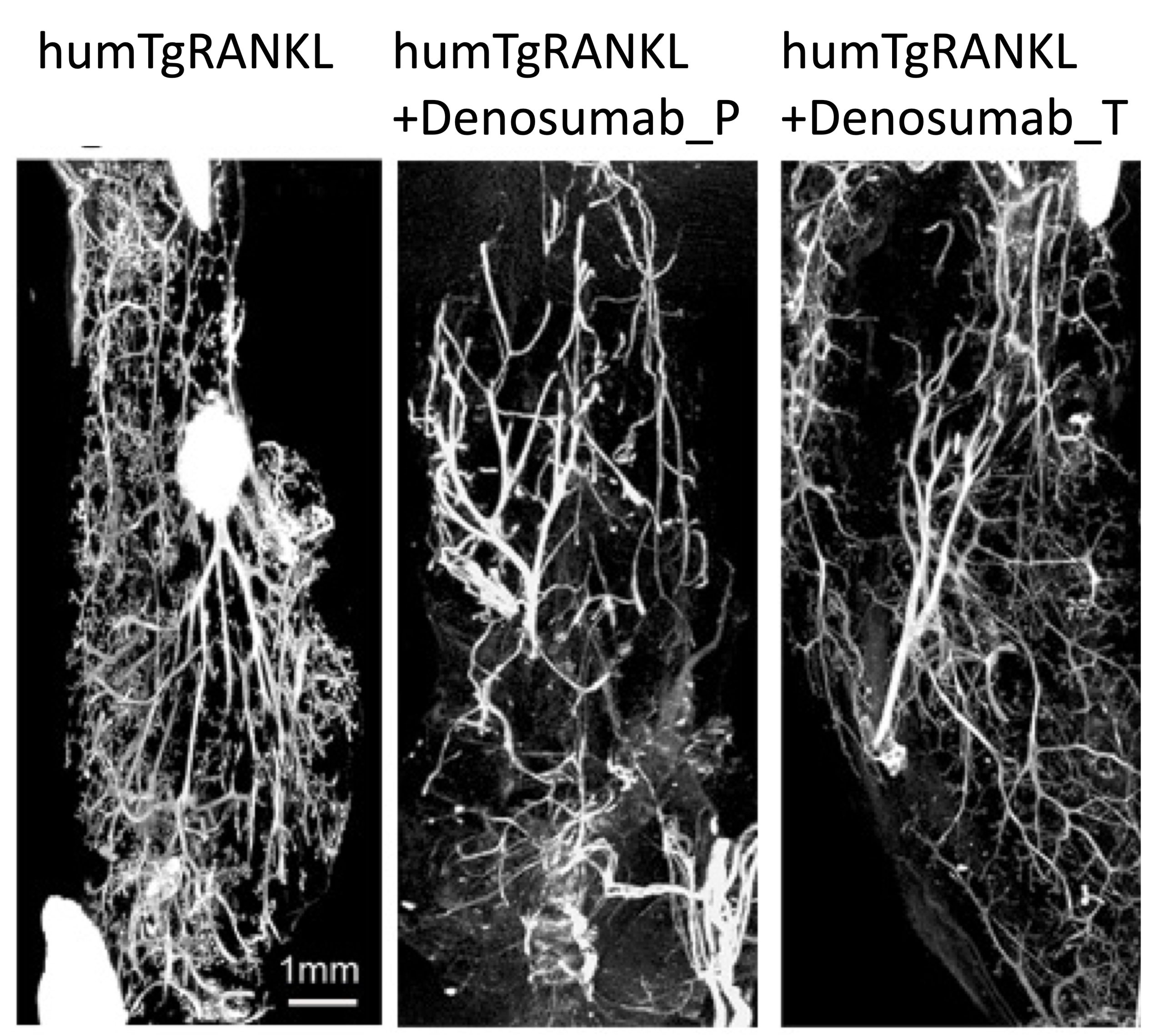
In the frame of a collaborative project co-financed by the European Union and Greek national funds (BreastCaRANKL project code: T1EDK-02829), Biomedcode has collaborated with Dr. Douni’s lab at BSRC Al. Fleming and the companies Bioemtech and Protavio to develop an innovative human-RANKL dependent breast cancer mouse model aiming to study RANKL dependent breast cancer mechanisms and to establish novel integrated preclinical platforms with modules of advanced imaging and molecular analysis for the evaluation of human therapeutics targeting cancer.
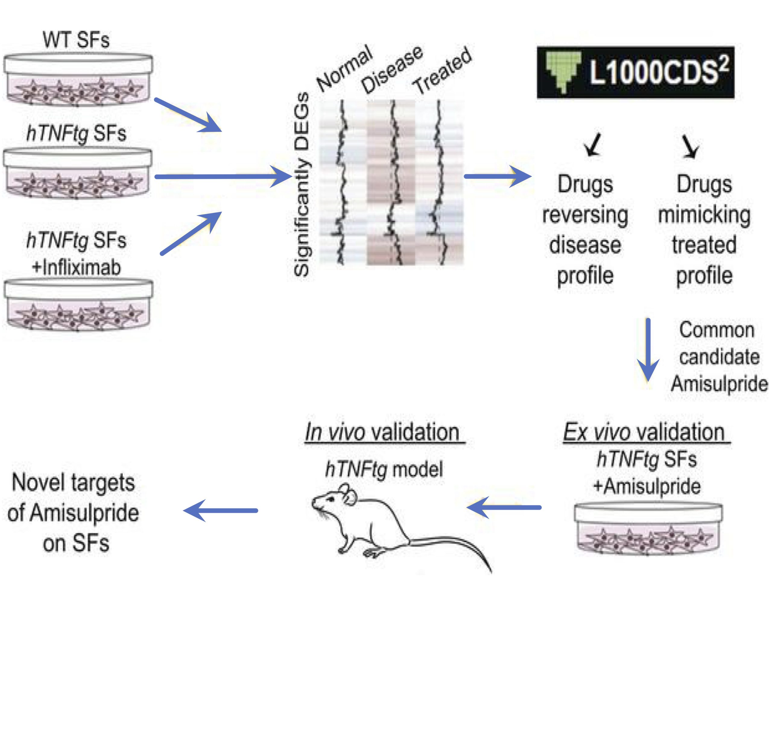
In the frame of a collaborative drug development project scientists from Biomedcode and BSRC Al. Fleming using bioinformatics tools, have repurposed the neuroleptic drug amisulpride for the reversal of the pathogenic expression signature of synovial fibroblasts and the treatment of arthritis pathology.
Published in JCI Insight 2023 May 8;8(9):e165024. doi: 10.1172/jci.insight.165024.
Cookies
To make this site work properly, we sometimes place small data files called cookies on your device. Most big websites do this too.
Accept
Change Settings
Cookie Box Settings
Cookie Box Settings
Privacy settings
Decide which cookies you want to allow.
You can change these settings at any time. However, this can result in some functions no longer being available. For information on deleting the cookies, please consult your browser’s help function.
Learn more about the cookies we use.
With the slider, you can enable or disable different types of cookies:
This website will:
Essential: Remember your cookie permission setting
Essential: Allow session cookies
Essential: Gather information you input into a contact forms, newsletter and other forms across all pages
Essential: Keep track of what you input in a shopping cart
Essential: Authenticate that you are logged into your user account
Essential: Remember language version you selected
This website won't:
Remember your login details
Functionality: Remember social media settings
Functionality: Remember selected region and country
Analytics: Keep track of your visited pages and interaction taken
Analytics: Keep track about your location and region based on your IP number
Analytics: Keep track of the time spent on each page
Analytics: Increase the data quality of the statistics functions
Advertising: Tailor information and advertising to your interests based on e.g. the content you have visited before. (Currently we do not use targeting or targeting cookies.
Advertising: Gather personally identifiable information such as name and location
This website will:
Essential: Remember your cookie permission setting
Essential: Allow session cookies
Essential: Gather information you input into a contact forms, newsletter and other forms across all pages
Essential: Keep track of what you input in a shopping cart
Essential: Authenticate that you are logged into your user account
Essential: Remember language version you selected
Functionality: Remember social media settings
Functionality: Remember selected region and country
This website won't:
Remember your login details
Analytics: Keep track of your visited pages and interaction taken
Analytics: Keep track about your location and region based on your IP number
Analytics: Keep track of the time spent on each page
Analytics: Increase the data quality of the statistics functions
Advertising: Tailor information and advertising to your interests based on e.g. the content you have visited before. (Currently we do not use targeting or targeting cookies.
Advertising: Gather personally identifiable information such as name and location
This website will:
Essential: Remember your cookie permission setting
Essential: Allow session cookies
Essential: Gather information you input into a contact forms, newsletter and other forms across all pages
Essential: Keep track of what you input in a shopping cart
Essential: Authenticate that you are logged into your user account
Essential: Remember language version you selected
Functionality: Remember social media settingsl Functionality: Remember selected region and country
Analytics: Keep track of your visited pages and interaction taken
Analytics: Keep track about your location and region based on your IP number
Analytics: Keep track of the time spent on each page
Analytics: Increase the data quality of the statistics functions
This website won't:
Remember your login details
Advertising: Use information for tailored advertising with third parties
Advertising: Allow you to connect to social sites
Advertising: Identify device you are using
Advertising: Gather personally identifiable information such as name and location
This website will:
Essential: Remember your cookie permission setting
Essential: Allow session cookies
Essential: Gather information you input into a contact forms, newsletter and other forms across all pages
Essential: Keep track of what you input in a shopping cart
Essential: Authenticate that you are logged into your user account
Essential: Remember language version you selected
Functionality: Remember social media settingsl Functionality: Remember selected region and country
Analytics: Keep track of your visited pages and interaction taken
Analytics: Keep track about your location and region based on your IP number
Analytics: Keep track of the time spent on each page
Analytics: Increase the data quality of the statistics functions
Advertising: Use information for tailored advertising with third parties
Advertising: Allow you to connect to social sitesl Advertising: Identify device you are using
Advertising: Gather personally identifiable information such as name and location
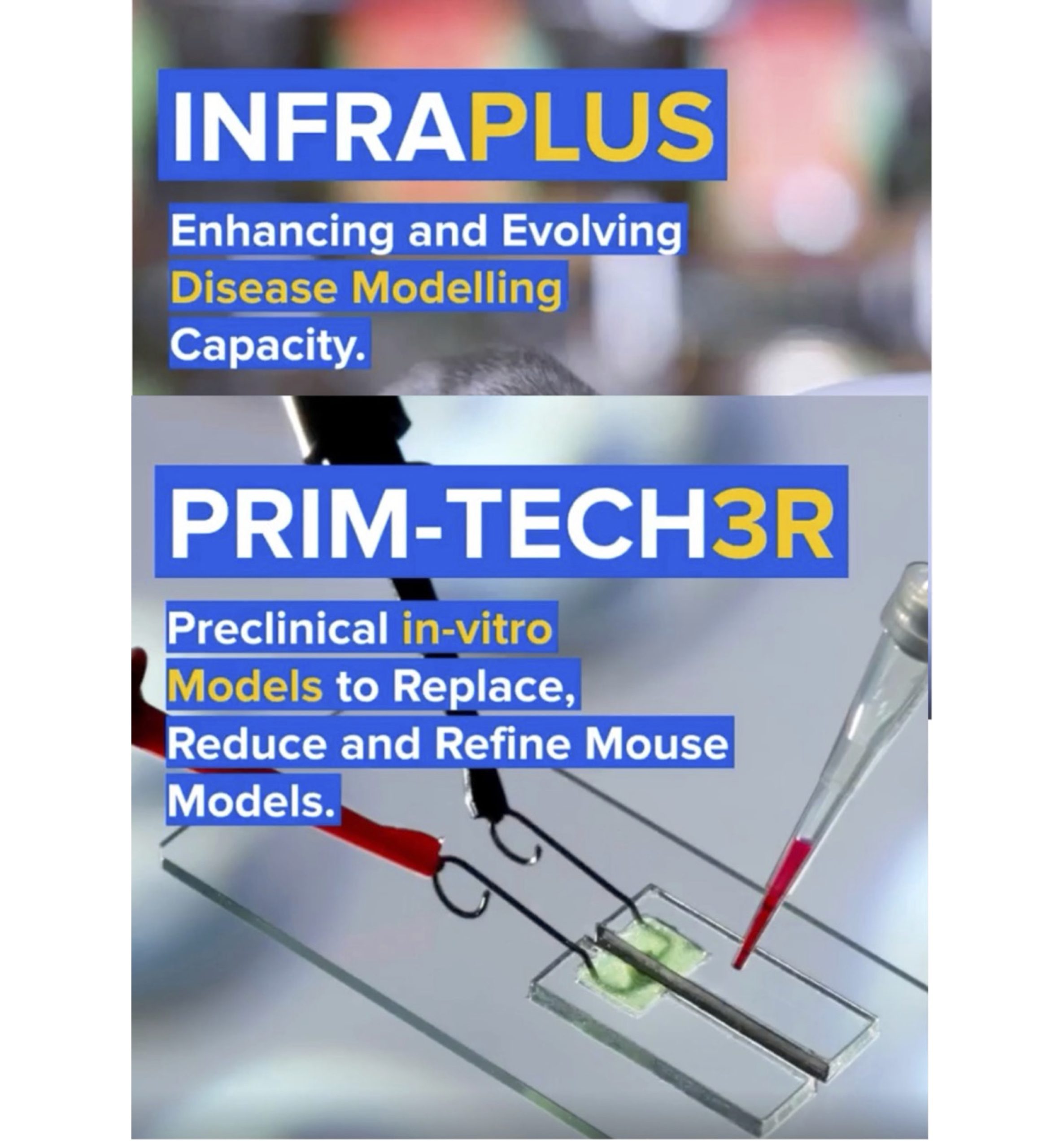

INFRAFRONTIER has achieved two major milestones in the first half of 2024, with two collaborative projects being funded by the European Commission INFRAPLUS and PRIMTECH3R
Biomedcode participates in both projects together with other partners and collaborators:
Helmholtz Munich – ERA-LEARN – Institut Clinique De La Souris ICS – CIPHE – Centre d’Immunophénomique – University of Oulu – Karolinska Institutet – CSIC – Centro Nacional de Biotecnología (CNB-CSIC) – Consiglio Nazionale delle Ricerche – Vetmeduni – BSRC Alexander Fleming – Universitat Autònoma de Barcelona – UK Research and Innovation – Mary Lyon Centre at MRC Harwell – MRC National Mouse Genetics Network – Biomedcode – AKITA, by Finnadvance – CNRS – Calouste Gulbenkian Foundation Instituto Gulbenkian de Ciencia – The Netherlands Cancer Institute – Antoni van Leeuwenhoek – The Hospital for Sick Children – Institute of Molecular Genetics of the Czech Academy of Sciences – Czech Centre for Phenogenomics – UMCG research – EMBL – INSERM and Aarhus University